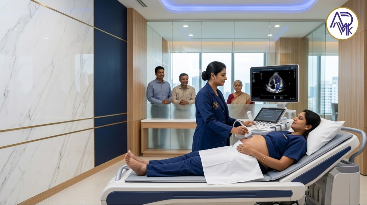
What Is a Fetal Echo Scan? An Exclusive Guide in 2026.
What Is a Fetal Echo Scan?
A Fetal Echo Scan, also known as Fetal Echocardiography, is a specialized ultrasound test used to examine the structure and function of a baby's heart before birth. It provides detailed images of the fetal heart, allowing doctors to assess the heart chambers, valves, blood vessels, heart rhythm, and blood flow.
This non-invasive and painless scan is usually recommended when there is a higher risk of congenital heart defects (CHDs) or if an abnormality is suspected during a routine pregnancy ultrasound. It helps in the early detection of heart conditions, enabling doctors to plan appropriate care during pregnancy, delivery, and after the baby is born.
A fetal echo scan is typically performed between 18 and 24 weeks of pregnancy, although it may be done earlier or later if advised by your doctor. It is safe for both the mother and the baby, as it uses sound waves rather than radiation.
What Are the Benefits of a Fetal Echo Scan?
- Early Detection of Heart Defects: Helps identify congenital heart abnormalities before birth.
- Detailed Heart Assessment: Evaluates the structure, function, rhythm, and blood flow of the baby's heart.
- Timely Medical Planning: Allows doctors to prepare appropriate treatment and care if a heart condition is detected.
- Better Pregnancy Management: Helps healthcare providers monitor high-risk pregnancies more effectively.
- Improved Delivery Planning: Ensures the baby is delivered at a facility equipped to provide specialized cardiac care if needed.
- Parental Reassurance: Confirms normal heart development in most pregnancies, providing peace of mind.
- Better Health Outcomes: Early diagnosis and intervention can improve treatment success after birth.
What Are the Types of Fetal Echo Scan?
There are three main types of fetal echo scans, depending on the baby's condition and the information required:
- 2D Fetal Echocardiography: The most common type, used to examine the structure and function of the baby's heart.
- Doppler Fetal Echocardiography: Evaluates blood flow through the heart, valves, and major blood vessels to detect circulation abnormalities.
- Color Doppler Fetal Echocardiography: Uses color imaging to show the direction and speed of blood flow, helping identify congenital heart defects and valve problems more accurately.
Your doctor will recommend the most suitable type of fetal echo scan based on your pregnancy and the
Is the Fetal Echo Scan Important?
Yes, a fetal echo scan is an important prenatal test that provides a detailed evaluation of your baby's heart before birth. It helps detect congenital heart defects, irregular heart rhythms, and other cardiac abnormalities that may not be visible during a routine pregnancy ultrasound.
Early diagnosis allows doctors to monitor the baby's condition, plan appropriate treatment, and ensure the right medical care is available at the time of delivery if needed. A fetal echo scan is especially recommended for pregnancies with risk factors, such as a family history of heart defects, maternal diabetes, certain infections, or abnormal findings on a routine ultrasound.
Why Is a Fetal Echo Scan Done?
A fetal echo scan is performed to examine the structure and function of your baby's heart before birth. It helps detect congenital heart defects, abnormal heart rhythms, and other cardiac conditions that may not be visible during a routine pregnancy ultrasound.
Doctors may recommend a fetal echocardiography if there is a family history of heart defects, maternal diabetes, certain infections, IVF pregnancy, abnormal ultrasound findings, or other high-risk pregnancy factors. Early diagnosis allows doctors to plan appropriate care during pregnancy, delivery, and after the baby is born, improving the chances of timely treatment if needed.
When Is a Fetal Echo Scan Done?
A fetal echo scan is usually performed between 18 and 24 weeks of pregnancy, as this is the ideal time to examine the baby's heart in detail. In some cases, your doctor may recommend the scan earlier or later, depending on your pregnancy and medical history.
This scan is advised if there is a family history of congenital heart disease, abnormal findings on a routine pregnancy ultrasound, maternal diabetes, certain infections, IVF pregnancy, or other factors that may increase the risk of fetal heart problems. Early detection helps doctors plan appropriate care before and after birth.
In Which Week Is a Fetal Echo Scan Done?
A fetal echo scan is usually performed between 18 and 24 weeks of pregnancy, as this is the ideal time to examine the baby's heart in detail. In some high-risk pregnancies, the scan may be recommended as early as 16 weeks, while a follow-up scan may be advised later if needed.
Your doctor may recommend a fetal echocardiography if there is a family history of congenital heart disease, abnormal ultrasound findings, maternal diabetes, certain infections, or other pregnancy-related risk factors. The scan helps detect heart abnormalities early, allowing for better pregnancy planning and newborn care.
In Which Month Is a Fetal Echo Scan Done?
A Fetal Echo Scan is usually performed between 18 and 24 weeks of pregnancy, which is around the 5th to 6th month. During this stage, the baby's heart is developed enough for the doctor to examine its structure, chambers, valves, and blood flow in detail.
In some high-risk pregnancies, your doctor may recommend a fetal echocardiography earlier or schedule follow-up scans later to monitor the baby's heart development. The exact timing depends on your medical history and your healthcare provider's advice.
Who May Need a Fetal Echo Scan?
A fetal echo scan may be recommended for pregnant women who have a higher risk of the baby developing a heart condition.
Your doctor may advise this scan if:
- There is a family history of congenital heart disease.
- The mother has diabetes, lupus, or certain infections during pregnancy.
- The pregnancy involves twins or more than one baby.
- An abnormality is detected on a routine pregnancy ultrasound.
- The baby has genetic or chromosomal conditions linked to heart defects.
- The mother has taken certain medications that may affect the baby's heart.
A fetal echo scan helps doctors assess the baby's heart in detail, allowing for early diagnosis and better pregnancy and delivery planning if a heart condition is detected.
How Is a Fetal Echo Scan Done?
A fetal echo scan is a safe, non-invasive ultrasound that examines your baby's heart before birth. During the scan, you will lie comfortably on an examination table while a clear gel is applied to your abdomen. The sonographer gently moves an ultrasound probe over your belly to capture detailed images of the baby's heart, including its chambers, valves, blood vessels, and blood flow.
The procedure is painless and usually takes 30 to 60 minutes, depending on the baby's position and the level of detail required. No needles, injections, or radiation are involved, making it safe for both the mother and the baby. After the scan, a specialist reviews the images and shares the findings with your doctor.
Are There Limitations to a Fetal Echo Scan?
Yes, a fetal echo scan has some limitations. While it provides a detailed assessment of your baby's heart, it may not detect every heart condition, especially very small defects or problems that develop later in pregnancy or after birth. Factors such as the baby's position, gestational age, maternal body type, and image quality can also affect the accuracy of the scan. Your doctor may recommend additional scans or follow-up evaluations if needed.
How to Prepare for a Fetal Echo Scan?
Preparing for a fetal echo scan is simple and usually requires no special preparation. You can eat and drink normally before the appointment unless your doctor advises otherwise. Wear comfortable, loose-fitting clothing to make the examination easier.
Bring your previous ultrasound reports, medical records, and referral documents, if available, as they help the specialist assess your baby's heart more accurately. During the scan, you will be asked to lie comfortably. At the same time, a gel is applied to your abdomen, and the procedure typically takes 30 to 60 minutes, depending on the baby's position and the complexity of the examination.
Try to arrive a few minutes early and stay relaxed, as a calm and comfortable experience helps ensure the best possible imaging results.
What Will the Fetal Echo Scan Results Show?
A Fetal Echo Scan provides detailed images of your baby's heart, allowing the doctor to evaluate its structure, function, heart chambers, valves, blood flow, and heartbeat. The scan can help detect congenital heart defects, abnormal heart rhythms (arrhythmias), and other cardiac abnormalities before birth. Early diagnosis enables doctors to plan appropriate care, treatment, and delivery if needed, ensuring the best possible outcome for both mother and baby.
What Are the Risks of Getting a Fetal Echo Scan?
A fetal echo scan is a safe, non-invasive ultrasound test that uses sound waves to examine your baby's heart. It does not use radiation, making it safe for both the mother and the baby when performed by trained healthcare professionals.
There are no known risks or harmful side effects associated with a fetal echocardiography scan. The procedure is painless, and you can resume your normal activities immediately after the test. The benefits of early detection of congenital heart conditions far outweigh any potential concerns.
A Fetal Echo Scan Is Indicated – What Happens Next?
If a fetal echo scan is indicated, your doctor will schedule the test with a specialist trained in fetal heart imaging. During the scan, detailed ultrasound images of your baby's heart are examined to check its structure, rhythm, and blood flow. If any abnormality is detected, your healthcare team will explain the findings, discuss possible treatment options, and plan appropriate care during pregnancy and after birth if needed. In many cases, the scan provides reassurance that your baby's heart is developing normally.
What Is the Fetal Echo Scan Price?
The price of a Fetal Echo Scan in India typically ranges from ₹2,500 to ₹6,000, depending on the city, diagnostic centre, doctor's expertise, and the technology used. In metro cities or advanced fetal medicine centres, the cost may be slightly higher.
Since prices can vary, it is best to contact your preferred diagnostic centre for the latest Fetal Echo Scan cost and to check if an appointment or doctor's referral is required.
Where Can I Get a Fetal Echo Scan Near Me?
If you're searching for a fetal echo scan near me, choose a trusted diagnostic centre or hospital with experienced fetal medicine specialists and advanced ultrasound technology. A fetal echocardiography scan requires specialized expertise to assess your baby's heart structure and function accurately.
- Experienced radiologists or fetal medicine experts
- Modern, high-resolution ultrasound equipment
- Accurate and timely reports
- Comfortable and hygienic facilities
- Easy appointment scheduling
A reliable diagnostic centre ensures accurate results, helping your doctor detect congenital heart conditions early and plan appropriate care for both mother and baby. Always book your scan at a centre recommended by your healthcare provider for the best possible care.
Conclusion
A Fetal Echo Scan is a safe, non-invasive, and highly specialized ultrasound that evaluates your baby's heart before birth. It plays a vital role in the early detection of congenital heart defects, allowing doctors to diagnose heart conditions, monitor fetal heart health, and plan the best possible care during pregnancy and after delivery.
If your doctor recommends a fetal echo scan, having it done at a trusted diagnostic centre staffed by experienced specialists can provide accurate results and peace of mind. Early diagnosis helps ensure timely medical care, supporting the health and well-being of both the mother and the baby.
Leave a comments