What is an HRCT thorax test? An Exclusive Guide in 2026.
What is an HRCT thorax test (high-resolution computed tomography) scan?
An HRCT thorax test (High-Resolution Computed Tomography of the chest) is an advanced imaging scan used to get very detailed images of the lungs and chest structures. It is especially designed to detect fine lung abnormalities that may not be visible on a normal chest X-ray or even a standard CT scan.Unlike a regular CT scan, an HRCT uses thin-slice imaging and high-resolution algorithms, allowing doctors to clearly see the airways, lung tissue, and interstitial spaces. This makes it one of the most accurate tests for diagnosing lung diseases at an early stage.
What is the HRCT thorax test used for?
The (High-Resolution Computed Tomography) HRCT Thorax test is primarily used to get highly detailed images of the lungs and chest structures. Unlike a routine chest X-ray or standard CT scan, HRCT focuses on fine lung details, making it one of the most accurate imaging tests for diagnosing lung and interstitial diseases.
Detect interstitial lung diseases (pulmonary fibrosis, sarcoidosis)
Evaluate chronic cough or breathlessness
Identify infections like TB or post-COVID lung damage
Diagnose COPD, bronchiectasis, and emphysema
Monitor lung disease progression and treatment response
Detect early lung nodules or abnormalities
What is the difference between an HRCT thorax test and a CT scan?
An (High-Resolution Computed Tomography) HRCT thorax test and a CT scan both use X-ray technology to create detailed images of the chest, but they are designed for different diagnostic purposes. Understanding the difference helps patients know why a doctor may recommend one over the other.
The main difference between an HRCT thorax test and a CT scan is the level of detail and purpose.
A CT scan is a general chest imaging test used to examine the lungs, heart, blood vessels, bones, and lymph nodes. It helps detect tumors, infections, injuries, and other chest conditions.
An HRCT thorax test is a specialized scan focused on the lungs. It uses very thin slices to detect early or subtle lung diseases such as pulmonary fibrosis, interstitial lung disease (ILD), chronic infections, and post-COVID lung changes.
Key Differences at a Glance
Focus: CT scan = whole chest | HRCT = lung tissue
Image detail: CT = standard | HRCT = very high resolution
Contrast: CT often uses contrast | HRCT usually does not
Best for: CT = tumors & trauma | HRCT = detailed lung evaluation
What are the benefits of an HRCT thorax test?
A (High-Resolution Computed Tomography) HRCT thorax test is an advanced imaging scan specially designed to give highly detailed images of the lungs and chest structures. Compared to a routine chest X-ray or standard CT scan, HRCT provides much clearer and more precise information, which makes it extremely valuable in diagnosing and managing lung diseases.
Early detection of lung diseases like fibrosis, ILD, infections, and COPD
Clear, high-resolution images that show small lung abnormalities
Accurate diagnosis of chronic and complex respiratory conditions
Helps differentiate between similar lung problems
Monitors disease progression and treatment response
Useful after lung infections to assess residual damage
Non-invasive, quick, and painless procedure
How do I prepare for an HRCT thorax test?
Preparing for a (High-Resolution Computed Tomography) HRCT Thorax scan is simple and usually stress-free. Proper preparation helps ensure clear images, accurate diagnosis, and a smooth scanning experience. Here’s everything you need to know before your test.
Preparing for an HRCT thorax scan is simple and usually requires minimal steps.
Fasting: Usually not required. If contrast is used, you may be asked to fast for 4–6 hours.
Medical History: Inform the doctor about allergies, kidney problems, diabetes, asthma, implants, or pregnancy.
Clothing: Wear loose, metal-free clothing. Avoid jewelry and metal accessories.
Medications: Continue regular medicines unless your doctor advises otherwise.
During the Scan: The test is quick, painless, and may require holding your breath briefly.
After the Scan: Resume normal activities. Drink plenty of water if contrast was used.
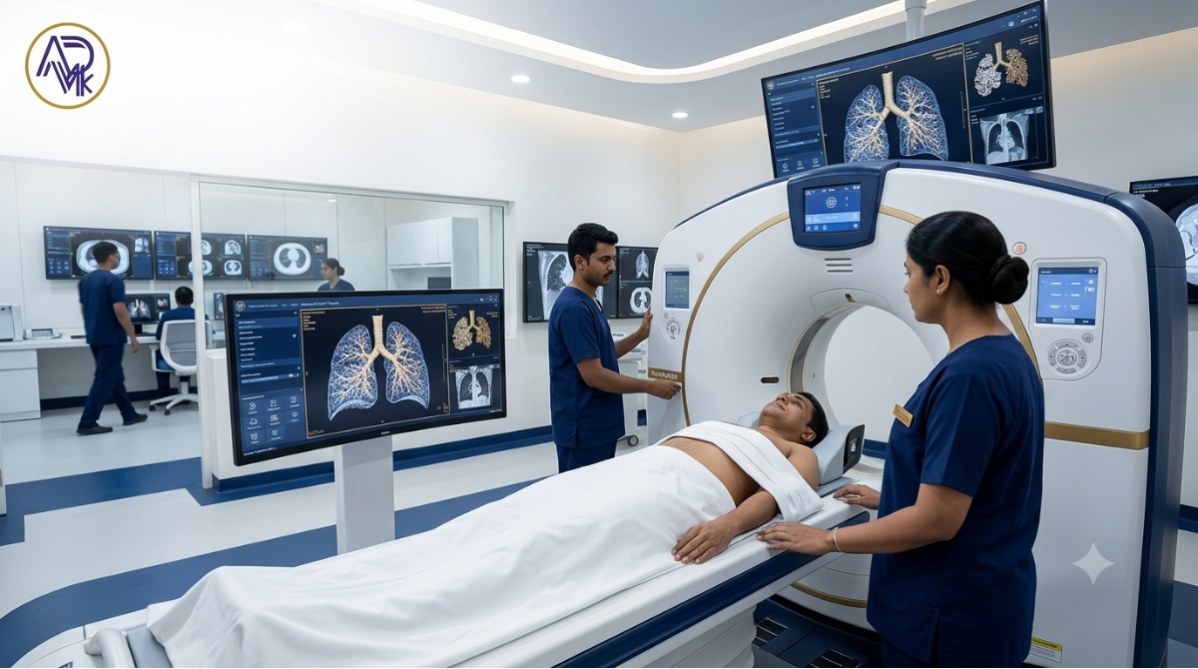
What happens during an HRCT thorax test?
A (High-Resolution Computed Tomography) HRCT thorax test is a painless, non-invasive imaging procedure used to get highly detailed images of the lungs and chest structures. Here’s a clear, step-by-step explanation of what typically happens during the test, written for patients and caregivers.
An HRCT thorax test is a quick, painless scan that takes detailed images of your lungs.
You lie flat on a CT table, usually with your arms above your head.
The table moves through a donut-shaped scanner.
You may be asked to hold your breath for a few seconds while images are taken.
The scan takes about 5–10 minutes and does not cause pain.
Contrast dye is usually not required, unless advised by your doctor.
How long does an HRCT thorax test take?
A (High-Resolution Computed Tomography) HRCT thorax test is a quick and efficient diagnostic scan designed to capture highly detailed images of the lungs and chest structures. Most patients are relieved to know that the test does not take much time and is usually completed within minutes.
An HRCT thorax test is quick and painless.
Actual scan time: 5–10 minutes
Total time at the center: Around 20–30 minutes (including registration and positioning)
After the scan, You can return to normal activities immediately
The scan itself is very fast, and in most cases, no contrast dye is required. Reports are usually available within 24 hours.
What happens after an HRCT thorax test?
After a (High-Resolution Computed Tomography) HRCT thorax test, the process is simple, safe, and usually stress-free for the patient. Here’s exactly what happens step by step, explained in clear, patient-friendly language:
The scan images are reviewed by a radiologist, who analyzes detailed views of the lungs and chest. A report is prepared highlighting any abnormalities such as infection, inflammation, scarring, or interstitial lung disease.
Your doctor then reviews the report with you and decides the next steps, which may include treatment, follow-up tests, or reassurance if results are normal.
What are the advantages of an HRCT thorax test?
A High-Resolution Computed Tomography) HRCT thorax test is an advanced imaging scan mainly used to examine the lungs and chest structures in great detail. Compared to a regular chest X-ray or standard CT scan, HRCT offers several important advantages, especially for diagnosing lung-related conditions early and accurately.
High-resolution imaging for clear, detailed lung views
Early detection of lung diseases like fibrosis, COPD, and infections
More accurate diagnosis compared to X-rays or regular CT scans
Better evaluation of subtle lung abnormalities
Helps monitor disease progression and treatment response
Non-invasive, painless, and quick procedure
Useful for assessing post-infection lung damage
What are the disadvantages of the HRCT thorax test?
A (High-Resolution Computed Tomography) HRCT thorax test is an advanced imaging test used to evaluate lung and chest conditions in great detail. While it is extremely useful for accurate diagnosis, it also has certain disadvantages and limitations that patients should be aware of before undergoing the test.
The HRCT thorax test is highly accurate for lung diagnosis, but it has some limitations:
Radiation Exposure: Uses X-rays, with higher radiation than a chest X-ray.
Not Safe in Pregnancy: Generally avoided due to radiation risk to the fetus.
Higher Cost: More expensive than X-rays and basic CT scans.
Limited Scope: Mainly evaluates lung diseases, not heart or other organs.
Patient Discomfort: Requires breath-holding and staying still.
Incidental Findings: May detect harmless abnormalities, causing anxiety.
Not for Routine Screening: Should only be done when medically required.
Why this HRCT thorax test?
A (High-Resolution Computed Tomography) HRCT Thorax test is done to get very detailed images of the lungs and chest structures. Doctors recommend this test when a regular chest X-ray or standard CT scan does not give enough clarity to diagnose a lung problem accurately.
Unlike routine scans, HRCT focuses on fine lung details, making it extremely useful for detecting early, subtle, or complex lung diseases.
Find the exact cause of breathing problems
Detect early lung diseases
Diagnose interstitial lung disease (ILD), pulmonary fibrosis, and sarcoidosis
Assess lung infections like TB, pneumonia, or COVID-related damage
Evaluate chronic conditions such as COPD and bronchiectasis
Check lung scarring or fibrosis
Monitor treatment progress
When and Who Needs to take an HRCT thorax test?
A (High-Resolution Computed Tomography) HRCT thorax test is a specialized imaging scan mainly used to get very detailed images of the lungs and chest structures. Doctors recommend this test when a routine X-ray or standard CT scan is not enough to clearly diagnose a lung or chest condition.
When Is an HRCT Thorax Test Needed?
Persistent cough or shortness of breath
Suspected lung diseases (ILD, fibrosis, COPD, bronchiectasis)
After severe lung infections (pneumonia, TB)
Abnormal chest X-ray or CT findings
To monitor lung disease progression or treatment response
Who Should Take an HRCT Thorax Test?
Patients with long-term breathing problems
Smokers or ex-smokers with respiratory symptoms
People exposed to dust, chemicals, or industrial fumes
Patients with autoimmune or systemic diseases
Elderly patients with declining lung function
What is the Best Time to Take the HRCT Thorax test?
The best time to take an (High-Resolution Computed Tomography) HRCT Thorax test depends mainly on your symptoms, doctor’s advice, and clinical condition, rather than the time of day. However, certain situations and timings can make the scan more effective and accurate.
HRCT gives the most accurate results during active symptoms
It can be done any time of the day
No fasting is required (unless contrast is used)
Early testing helps in faster diagnosis and better treatment decisions
What are the eligibility criteria for the HRCT thorax test?
The (High-Resolution Computed Tomography) HRCT thorax test is a specialised imaging scan of the lungs and chest used to detect detailed structural changes that often aren’t visible on regular X-rays or standard CT scans. It’s not a routine test for everyone — doctors recommend HRCT only when certain clinical criteria are met.
You may be eligible if you have:
Persistent cough or shortness of breath
Unexplained chest symptoms
Suspected interstitial lung disease (ILD)
Ongoing symptoms after severe lung infection or COVID-19
Abnormal findings on chest X-ray or lung function tests
What is the list of Parameters for the HRCT thorax test?
The (High-Resolution Computed Tomography) HRCT Thorax test evaluates the lungs and surrounding chest structures in very fine detail. Unlike a routine chest CT, HRCT focuses on microscopic lung patterns, making it the gold standard for diagnosing interstitial and chronic lung diseases.
Doctors analyze multiple specific parameters in an HRCT thorax scan to accurately identify abnormalities, disease severity, and progression.
Below is a comprehensive list of HRCT thorax parameters commonly assessed by radiologists.
1. Lung Parenchyma
Ground-glass opacities
Consolidation
Fibrosis & honeycombing
Nodules
Air trapping
Emphysema
2. Airways
Bronchiectasis
Bronchial wall thickening
Airway narrowing
Mucus plugging
3. Lung Distribution
Upper or lower lobe involvement
Central or peripheral disease
Diffuse or focal patterns
4. Pleura
Pleural effusion
Pleural thickening
Fibrotic changes
5. Lymph Nodes & Mediastinum
Hilar & mediastinal lymph nodes
Calcifications or masses
6. Vascular Findings
Pulmonary artery size
Signs of pulmonary hypertension
Why these Parameters are Important
These parameters help diagnose conditions like ILD, pulmonary fibrosis, COPD, infections, post-COVID lung damage, and TB, and guide treatment decisions.
What Are Illnesses Diagnosed with HRCT Without Contrast?
A (High-Resolution Computed Tomography) HRCT test of the chest without contrast is one of the most accurate imaging tests for evaluating lung and airway diseases. It provides extremely detailed images of lung tissue, making it especially useful for diagnosing conditions that may not appear clearly on X-rays or routine CT scans.
Interstitial Lung Diseases (ILDs) – pulmonary fibrosis, sarcoidosis, hypersensitivity pneumonitis
COVID-19 & post-COVID lung changes – ground-glass opacities, fibrosis
COPD & emphysema – air trapping, bullae
Bronchiectasis – dilated airways, mucus plugging
Tuberculosis (TB) – active TB, old scars, cavities
Lung infections – bacterial, viral, fungal pneumonia
Pulmonary nodules – small benign or suspicious lung nodules
Occupational lung diseases – silicosis, asbestosis
What is the Procedure for Taking an HRCT Thorax Test?
A (High-Resolution Computed Tomography of the chest) HRCT Thorax test is a non-invasive imaging test used to get highly detailed images of the lungs and chest structures.
Before the Scan
Usually no special preparation is needed
Remove metal objects and wear comfortable clothing
Inform the doctor about allergies, pregnancy, or kidney issues (if contrast is used)
During the Scan
You lie on a table that slides into the CT scanner
You may be asked to hold your breath briefly
The scan takes 5–10 minutes and is completely painless
After the Scan
Normal activities can be resumed immediately
Drink fluids if contrast was used
What are the Risks & Limitations for the HRCT thorax test?
A (High-Resolution Computed Tomography) HRCT thorax test is a highly advanced imaging test used to detect detailed lung and chest abnormalities. While it is extremely useful for diagnosis, patients should also understand its risks and limitations before undergoing the test.
Risks
Radiation exposure (higher than X-ray, but low and controlled)
Not advised during pregnancy unless absolutely necessary
Contrast dye risks (rare allergies or kidney issues in susceptible patients)
Mild discomfort or claustrophobia in some individuals
Limitations
Shows structural changes only, not lung function
Cannot replace clinical examination or blood tests
May detect minor, non-serious findings leading to extra tests
Costlier than basic imaging like chest X-rays
In which case is the HRCT thorax test not performed?
A (High-Resolution Computed Tomography) HRCT thorax test is a powerful diagnostic tool for lung and chest conditions, but it is not recommended or avoided in certain situations due to safety concerns, limited clinical value, or better alternative tests being available.
Below are the key cases where an HRCT thorax test is usually not performed or is done only with strict medical justification:
Pregnancy, especially in the first trimester (due to radiation risk)
Children, unless necessary
Routine checkups or screening without symptoms
When a chest X-ray is sufficient for diagnosis
Patients who cannot stay still or hold their breath
Severe kidney disease (if contrast is required)
Patients with multiple recent CT scans (to avoid excess radiation)
When other tests are more suitable (e.g., MRI, ultrasound, lung function tests)
When should I call my healthcare provider?
You should contact your healthcare provider if you are experiencing symptoms that are persistent, worsening, or unusual, especially when they involve your lungs or breathing. Early communication helps in timely diagnosis and prevents complications.
Contact your doctor if you experience:
Persistent shortness of breath
Chronic cough or chest discomfort
Fever that doesn’t improve
Coughing up blood
Repeated respiratory infections
Seek urgent medical help if you have:
Sudden or severe breathing difficulty
Sharp chest pain
Bluish lips or fingertips
What is the HRCT Thorax Test Price in India?
The cost of a (High-Resolution Computed Tomography) HRCT Thorax test in India can vary significantly depending on the city, type of facility (private vs government), equipment used, and whether contrast is required.
The HRCT Thorax test price in India typically ranges between ₹3,000 to ₹6,000 for a plain scan at most private diagnostic centres. If contrast is required, the cost may increase to ₹4,500–₹9,000. Prices vary based on the city, diagnostic centre, scan technology, and use of contrast. Government hospitals usually offer the test at a lower, subsidized cost.
How to find an HRCT Chest-Contrast Centre near me in Delhi?
Here’s how you can find an HRCT Chest-Contrast centre near you in Delhi, plus a list of trusted diagnostic centres that offer HRCT / Chest CT & contrast scans 👇
Finding an HRCT Chest-Contrast scan centre in Delhi is simple if you follow these steps:
Search on Google or Google Maps using keywords like “HRCT Chest-Contrast centre near me in Delhi” to see nearby options.
Check diagnostic lab websites or healthcare platforms to compare services, prices, and reviews.
Call the centre before visiting to confirm availability of HRCT with contrast, appointment timing, cost, and preparation.
Carry a doctor’s prescription, as contrast scans usually require one.
Ask about preparation, such as fasting and recent kidney function test reports.
Conclusion
An HRCT thorax test (High-Resolution Computed Tomography scan) is a highly advanced imaging test that provides clear, detailed views of the lungs and chest structures. It plays a crucial role in early detection, accurate diagnosis, and monitoring of lung diseases, especially conditions that may not be visible on a normal X-ray or standard CT scan.
Because HRCT uses thin-slice imaging, doctors can identify subtle abnormalities such as interstitial lung disease, fibrosis, infections, or post-COVID lung changes with greater precision. The test is non-invasive, quick, and generally safe when performed under medical guidance.
If your doctor has advised an HRCT thorax scan, it is usually to gain deeper insight into your lung health and plan the most effective treatment. Always discuss your results with a qualified healthcare professional to understand what they mean for your condition and next steps.
Leave a comments